What It Is
A bone density test, most commonly performed using a technique called dual-energy X-ray absorptiometry, or DEXA, measures the density of minerals, primarily calcium, within bone tissue.
What It Checks
The test measures how much bone mineral is present within a specific area of bone, most commonly at the hip and spine, which are frequently used as representative sites for assessing overall bone strength throughout the skeleton.
Why It's Used
Bone density testing is commonly used to assess overall bone health, particularly relevant to conditions involving bone strength that can develop with age or other contributing factors, and is often used to track changes in bone density over time.
How It's Done
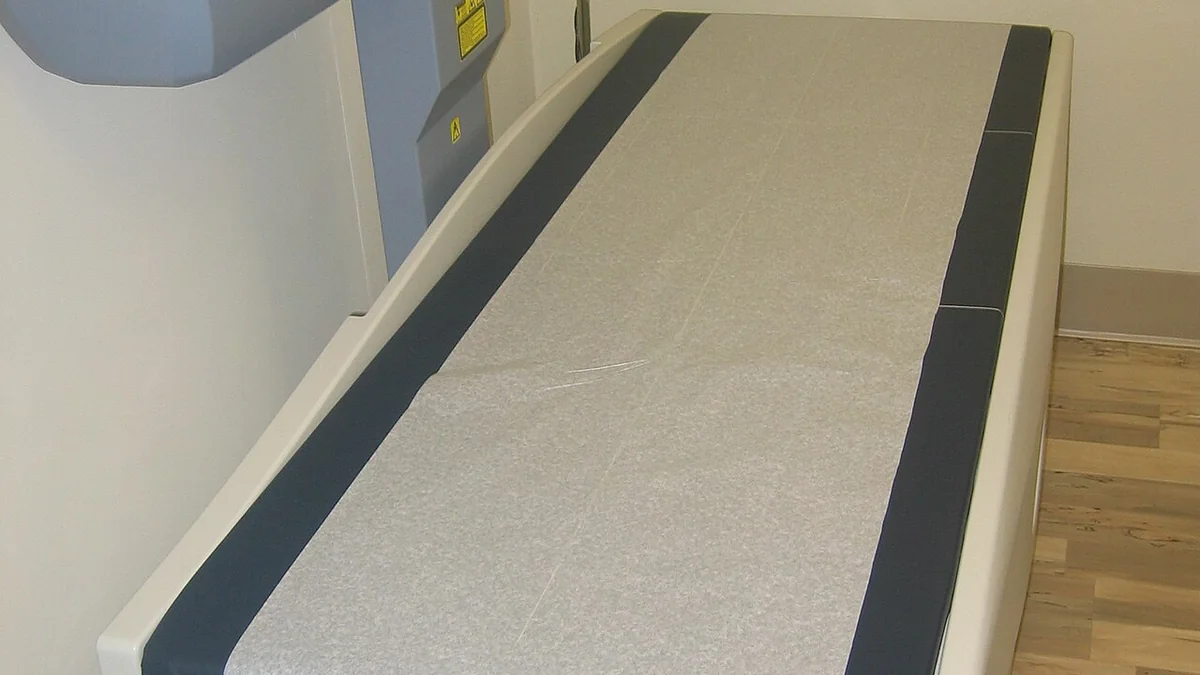
During a DEXA scan, the person lies still on a table while a scanning arm passes over the body, sending a very low dose of X-rays through the targeted bones. The amount of X-ray energy absorbed by the bone is used to calculate its mineral density. The procedure is quick, generally taking only about ten to twenty minutes, and involves a much lower radiation dose than a standard X-ray.
Background
Earlier methods for assessing bone density were developed in the mid-20th century but had notable limitations in accuracy and consistency. Dual-energy X-ray absorptiometry was developed in the late 1980s, offering a more precise and standardized way to measure bone mineral density, and it quickly became the preferred method for bone density assessment. It remains the most widely used and internationally recognized standard for bone density testing in healthcare today.