What It Is
A computed tomography scan, commonly known as a CT scan, is an imaging test that uses X-rays taken from multiple angles to create detailed cross-sectional images of the inside of the body.
What It Checks
A CT scan can produce detailed images of a wide range of structures, including bones, blood vessels, and organs throughout the body. Because it captures images from many angles and combines them, it can reveal more detail and depth than a standard X-ray, allowing for a closer look at the size, shape, and position of internal structures.
Why It's Used
CT scans are commonly used to closely examine internal organs and structures, particularly in situations where more detail is needed than a standard X-ray can provide, and are used across a wide range of clinical evaluations given their speed and level of detail.
How It's Done
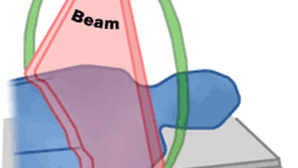
During a CT scan, the person lies still on a table that slowly moves through a large, ring-shaped scanner. The scanner rotates around the body, taking a series of X-ray images from different angles, which a computer then processes and combines into detailed cross-sectional, and sometimes three-dimensional, images. The procedure is generally quick, often taking just a few minutes.
Background
The mathematical principles needed to reconstruct cross-sectional images from multiple X-ray angles were developed earlier in the 20th century, but it wasn't until the early 1970s that the first practical CT scanner was built, a development that earned its inventors a Nobel Prize. Since then, CT technology has advanced considerably, becoming faster and capable of producing increasingly detailed images, and it's now one of the most widely used advanced imaging tools in modern medicine.