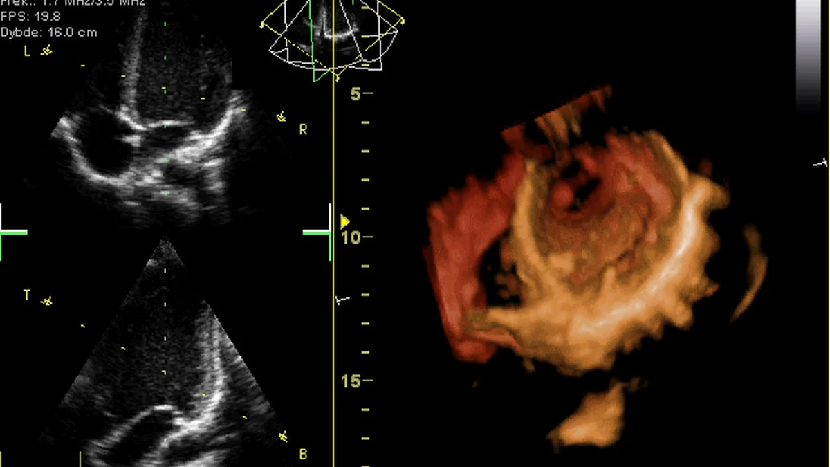
What It Is
An echocardiogram is an ultrasound-based imaging test focused specifically on the heart, producing real-time moving images of the heart as it beats.
What It Checks
An echocardiogram shows the heart's chambers, valves, and surrounding structures in motion, allowing a detailed look at how the heart is contracting, how blood is moving through its chambers, and how well its valves are opening and closing with each heartbeat.
Why It's Used
Because it captures the heart in real-time motion, an echocardiogram is commonly used to evaluate overall heart structure and function, investigate symptoms that might relate to the heart, and monitor known heart conditions over time.
How It's Done
An echocardiogram is typically performed by placing a small ultrasound transducer against the chest, in a similar way to a general abdominal ultrasound, using sound waves to generate moving images of the heart displayed on a screen in real time. The standard version of the test is done from outside the body and generally takes around thirty minutes to an hour to complete.
Background
Echocardiography developed from the same underlying sound wave imaging technology used in general ultrasound, with early experimental use for examining the heart beginning in the 1950s. The technology advanced considerably over subsequent decades, including the addition of color imaging to help visualize blood flow direction, further refining the level of detail available. Echocardiography is now one of the most widely used and valued tools for evaluating heart structure and function, given its ability to show the heart working in real time without radiation.